https://newatlas.com/3d-color-xrays-cern/55403/
I heartily approve of this. I wish I’d thought of it.


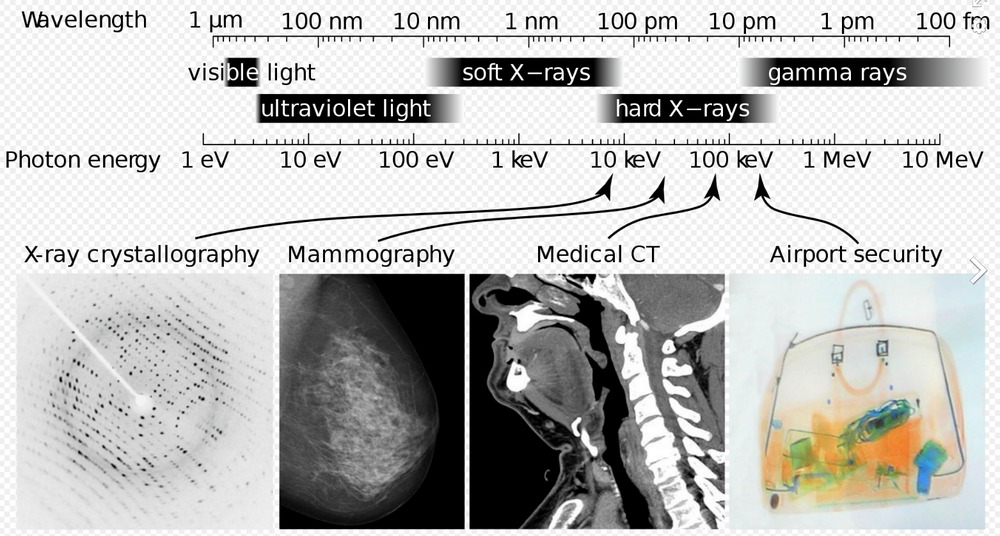
Medical X-ray scans have long been stuck in the black-and-white, silent-movie era. The contrast helps doctors spot breaks and fractures in bones, but more detail could help pinpoint other problems. Now, a company from New Zealand has developed a bioimaging scanner that can produce full color, three dimensional images of bones, lipids, and soft tissue, thanks to a sensor chip developed at CERN for use in the Large Hadron Collider. A 3D color image is generated that clearly shows muscle, bone, water, fat, disease markers – and even a watch. The end results are unnerving, like someone’s sculpted a detailed clay model of your insides.
At the heart of the Spectral CT scanner is a Medipix3 chip. This device, which detects and counts every individual particle that hits each pixel on the sensor, was originally developed at CERN to precisely track particles in the Large Hadron Collider. Measure the attenuation of specific wavelengths of the X-rays as they pass through different materials.