Michael V said:
Interesting, thanks. It’s extremely interesting that palynomorphs were preserved in the goethitic rocks. This is highly unusual. I’ll go read the original paper and see whether I can get to understand why.
https://www.science.org/doi/10.1126/sciadv.abm1406
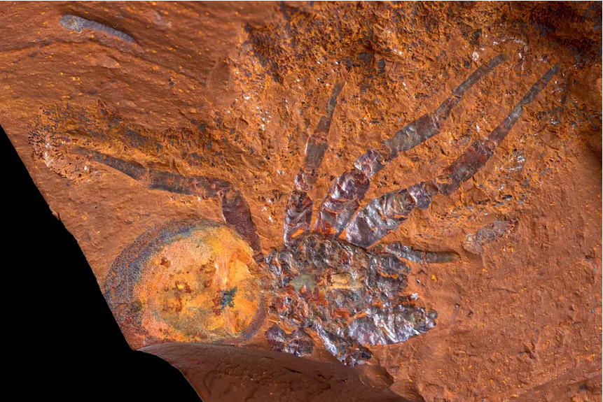
It turns out that these are replacement fossils; remains were directly replaced by goethite, likely very soon after burial. No organic material remains. The preservation is exquisite. Have a look at the size of the scale bars in the images below!
“The detailed preservation of microfossils in this deposit has implications for palynological studies of other goethite deposits, as it demonstrates that scanning electron microscopy (SEM) scanning of oxidized, iron-rich sediments (previously deemed unproductive using standard palynological techniques) can, under certain circumstances, enable the identification of microfossils and aid in the dating of sites.”
This new discovery has serious implications in the world of paleopalynology. Serious implications.
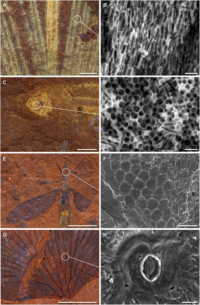
HIGH-FIDELITY PRESERVATION IN FOSSILS FROM MCGRATHS FLAT.
(A and B) Microstructure of fossil feather (AM F.145096) showing melanosome preservation; (C and D) melanosomes preserved in the eye of a fossil fish (AM F.145094); (E and F) crane fly (Limoniidae cf. Tonnoirella) fossil (AM F.146583) with ommatidia of the eye preserved; (G and H) Lygodium leaf (AM F.146600) exhibiting the structure of the stomata. Scale bars, 1 μm (B and D), 10 μm (H), 50 μm (F), 0.5 mm (A), and 5 mm (C, E, and G).
——————————————————————————————————————————————————————————————————————————————————
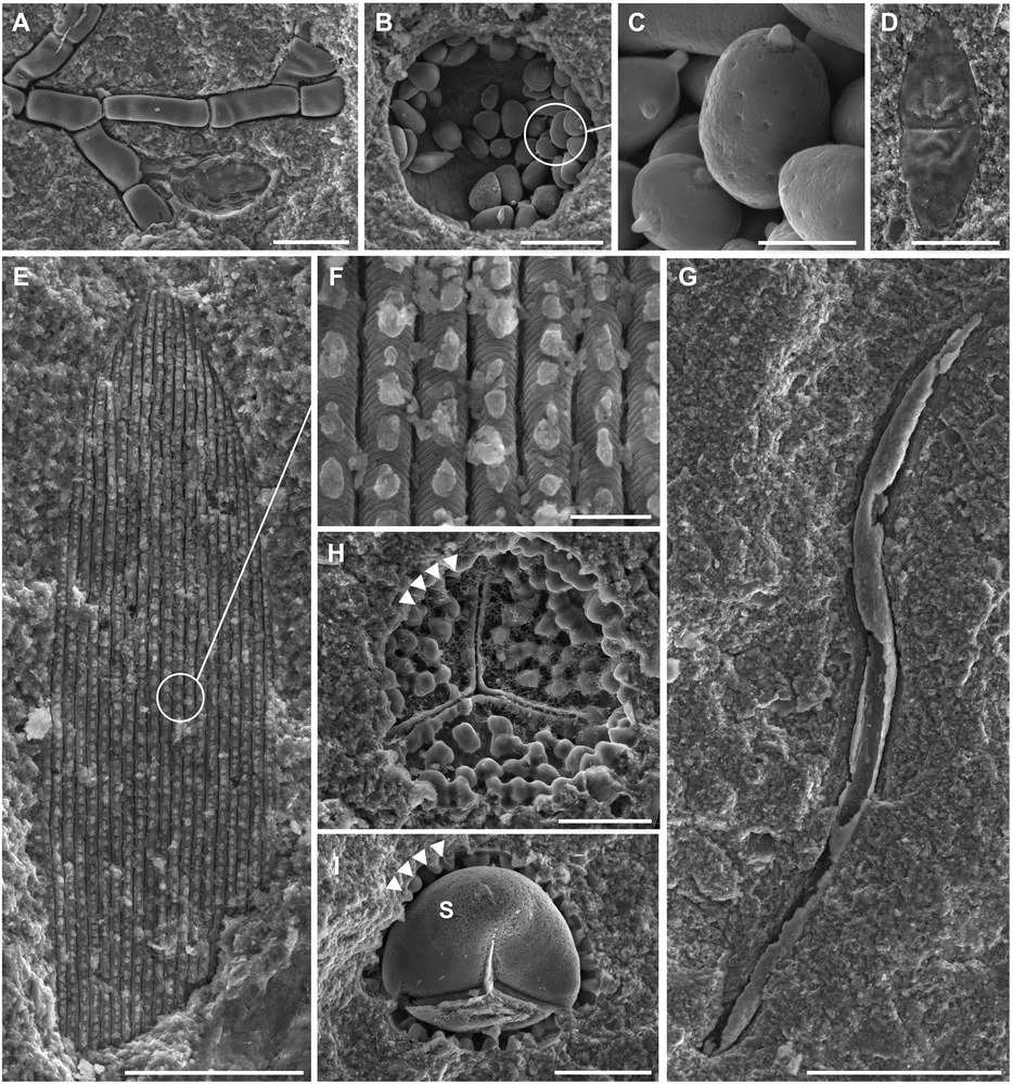
MISCELLANEOUS MICROFOSSILS.
(A) Fungal hypha (AM F.146156.2); (B and C) yeast-like fungal cells (AM F.145847.1); (D) Dicellasporites sp. (AM F.145716.1); (E and F) lepidopteran wing scale (AM F.146089.1); (G) nematode (AM F.145716.2); (H) Rugulatisporites trophus (AM F.145717.1) showing the imprint of exine (arrowheads); and (I) Rugulatisporites sp. with both exine (arrowheads) and intine steinkern (S) (AM F.146155.1). Scale bars, 2 μm (F), 5 μm (D), 10 μm (H and I), 20 μm (A, B, and E), and 50 μm (C and G).
——————————————————————————————————————————————————————————————————————————————————
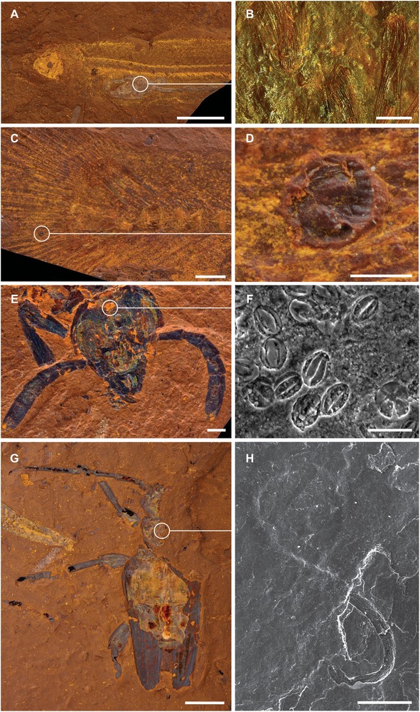
EVIDENCE OF BIOTIC INTERACTIONS.
(A and B) Stomach contents of fish (AM F.145094) showing that it fed predominantly on phantom midges (Chaoborus sp.); (C and D) a parasitic glochidium attached to the caudal fin of a fish (AM F.146607); (E and F) pollen preserved on the head of a sawfly (AM F.145093); and (G and H) a phoretic nematode attached to the body of a longhorn beetle (Cerambycidae: Coleoptera; AM F.145098). Scale bars, 25 μm (F), 100 μm (H), 250 μm (B and D), 1 mm (C and E), 5 mm (G), and 10 mm (A).
——————————————————————————————————————————————————————————————————————————————————