mollwollfumble said:
Ever heard of an X-ray microscope?
There are X-ray telescopes such as Chandra, they use multiple mirrors with grazing incidence reflection to focus the X-rays. (Wikipedia lists an amazing 52 X-ray telescopes in space). I haven’t heard of anything similar in microscopes.
Wikipedia has an article on X-ray microscopes, but it’s quite garbled because for instance the introduction directly conflicts with the section “Advanced Light Source”, etc. It’s also not clear from the article whether X-ray microscopes are portable like other microscopes or require a synchrotron for their X-ray source. Synchrotrons are not portable.
How much would an X-ray microscope cost and what could I do with it?
This one costs about $1.7 million USD.
The highest resolution—down to 50 nanometers—3D X-ray imaging available in a laboratory. Typically used for materials science, eg. porosity of rock or material failure in tension. The 3-D is tomography. The field of view from 16 to 65 µm, so the samples have to be really small. (Other machines have bigger field of view up to at least 100 mm across but coarser resolution, which can image a human knee for example). Energy at 5.4 keV, what’s that in wavelength? 0.23 nanometres, that’s very small, but it’s still considered a soft X-ray because hard x-rays have wavelengths shorter than 0.2 nm.
I haven’t yet seen any X-ray microscope that doesn’t include 3-D tomography.
But what about X-ray source and optics, very little mentioned on the web. There are at least three different types of X-ray focussing devices, as well as devices like a “diffuser” that spreads a point source.

This article is interesting. It’s about 2-D and 3-D X-ray microscopy of cells using a DIY benchtop design.
Affordable X-Ray Microscopy with Nanoscale Resolution
Transmission electron microscopy is limited to samples less than 500 nm thick, which is not thick enough for cells.
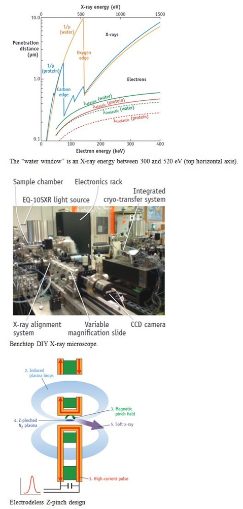
Soft x-ray tomography within the “water window” (that is, the wavelength range for which water is transparent to x-rays while other elements such as carbon and nitrogen absorb) enables direct imaging of biological specimens up to 10 μm thick, which is good for cells.
In the literature, one X-ray source makes use of a custom liquid-jet high-brightness laser-plasma to acquire images in less than 10 seconds, while another uses a commercially available Z-pinch source capable of a single image in 30 seconds.
Traditional Z-pinch plasma sources erode the electrodes which coats the optics, reducing system life. On the other hand, laser plasma sources, where a jet of liquid nitrogen is heated with pulses from a high-performance laser, last only a few hours. So the design chosen was an electrodeless Z-pinch design. Even so, it needs maintenance after 20 days of operation.